


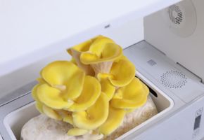
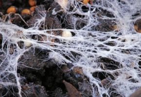

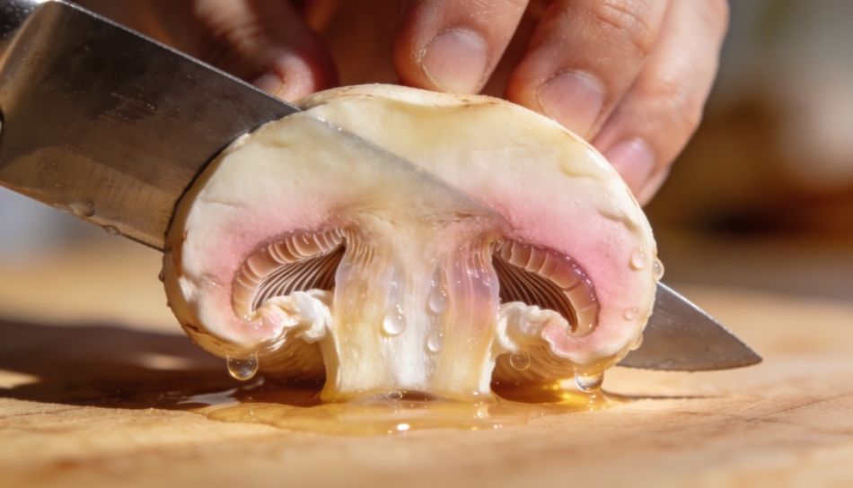
Have you ever cut a fresh mushroom and watched, amazed, as its white or yellowish flesh transformed within seconds into an intense blue, brick red, or raven black? This phenomenon, seemingly magical, has fascinated foragers, chefs, and naturalists for centuries. Yet it is one of the most elegant manifestations of fungal biochemistry: a chemical defense system, an evolutionary signal, an invaluable diagnostic indicator. Mushrooms change color for profound and scientifically documented reasons, involving enzyme chemistry, forest ecology, and, for those who cultivate or forage them, food safety. Understanding why mushrooms change color is not merely an intellectual curiosity exercise: it is a concrete skill that helps amateur mycologists identify species, growers monitor mycelium quality, chefs better preserve products, and researchers explore molecules of pharmacological interest. In this article, we will explore every facet of this phenomenon with the rigor it deserves, analyzing the chemistry of mushroom oxidation, the most interesting species, practical implications for foraging and cultivation, and aspects still open in scientific research. Whether you are an enthusiast who combs the undergrowth every autumn, a hobbyist growing oyster or shiitake mushrooms at home, a plant biology student, or a food service professional, this comprehensive guide will provide everything you need to know about the phenomenon of chromatic shifting in mushrooms. Prepare to see the fungal kingdom with completely new eyes. Chromatic shifting in mushrooms is one of the most visually spectacular phenomena observable in nature. When mushrooms change color following cutting, pressure, a bite, or simply due to the passage of time, they are telling us something precise about their internal biology. This is not an evolutionary whim nor an accessory characteristic: it is a highly coordinated biochemical response involving enzymes, signaling molecules, and secondary pigments. The technical term used by mycologists to describe this phenomenon is staining or, in English, bluing reaction when referring to the most famous case (blue staining in boletes). More generally, mycologists speak of mechanical damage response or oxidative response. In any case, the fundamental process underlying it is always the same: the rupture of cellular structures brings substances that are normally separated into contact, and their encounter in the presence of oxygen generates colored compounds. Chromatic shifting is much more common than one might think. An estimate published in 2018 in the Journal of Fungi suggests that over 15% of known macroscopic fungal species show some form of chromatic change upon injury. This percentage rises significantly if one considers more subtle color variations, such as slight browning or a shift to pale yellow visible only under optimal conditions. These numbers, however approximate, give us an idea of the scope of the phenomenon. It should be noted that research on fungal pigmentation is still incomplete: it is estimated that less than 10% of the world's fungal species has been described scientifically, and of these only a fraction has been analyzed from a biochemical point of view. For those who approach mushrooms with practical intentions (foraging for food consumption, home or professional cultivation, study for academic purposes), chromatic shifting has concrete and important implications. First of all, it is one of the most immediate and reliable diagnostic characteristics for identifying species in the field. Secondly, it reveals information about freshness and conservation status. Thirdly, it signals the presence of biologically active molecules that are increasingly interesting pharmacological and cosmetic research. An experienced forager knows, for example, that a bolete that stains immediately and intensely blue when cut must be identified with greater care: in the bolete family, rapid and intense staining can distinguish excellent edible species from toxic species with a similar appearance. However, it is not sufficient alone — as we will see — and must always be combined with all other morphological characteristics. At the base of all chromatic shifting phenomena is a precise biochemical process: enzymatic oxidation. To fully understand why mushrooms change color, it is necessary to take a step back and understand what oxidative enzymes are, which substrates they attack, and what products they generate. This knowledge is not only theoretical: it has direct applications in food preservation, industrial biotechnology, and the search for new drugs. The main enzymes involved in chromatic shifting of mushrooms belong to two large families: phenol oxidases and peroxidases. Let's examine them in detail. Laccases are copper-containing enzymes that catalyze the oxidation of a wide range of phenolic substrates using molecular oxygen directly as an electron acceptor. They are present in many fungal species (both Ascomycetes and Basidiomycetes) and perform fundamental functions in lignin degradation, pigment formation, and protection of the fungus from pathogens. In intact tissues, laccases are confined in cellular compartments separated from their substrates: only when the cell is damaged do the two components meet and the reaction begins. Scientific research has identified extraordinary biotechnological potential in fungal laccases. They are used in the textile industry for dye decolorization, in the paper industry for wood pulp delignification, in oenology for wine stabilization, and even in biosensor production. Fungal laccases are generally more stable and efficient than plant laccases, making them privileged objects of study. Tyrosinase, also known as bifunctional polyphenol oxidase, is a copper-containing enzyme that catalyzes two distinct reactions: the hydroxylation of L-tyrosine to L-DOPA (cresolase activity) and the oxidation of L-DOPA to dopaquinone (catecholase activity). Dopaquinone is highly reactive and spontaneously polymerizes forming dark melanins or reacts with other compounds to form colored pigments of various natures. Tyrosinase is the enzyme responsible for browning in many common mushrooms, including Agaricus bisporus (cultivated button mushroom) and Lentinula edodes (shiitake). Inhibition of tyrosinase is one of the main objectives of the food and cosmetic industries: in the food industry because enzymatic browning reduces the commercial quality of products, in the cosmetic field because melanin produced by tyrosinase is responsible for hyperpigmented skin spots. Peroxidases are enzymes that use hydrogen peroxide (H₂O₂) as an oxidant to catalyze the oxidation of various substrates. In fungi, the most studied are lignin peroxidase (LiP) and manganese peroxidase (MnP), mainly involved in lignin degradation in white rot fungi. However, some peroxidases also contribute to pigment formation at the time of tissue damage. Less relevant from the point of view of immediate staining compared to laccases and tyrosinases, peroxidases nevertheless play a crucial role in long-term oxidation processes and pigment stabilization. The enzymes mentioned attack specific substrates. In fungal staining chemistry, the protagonists are mainly phenolic compounds: molecules characterized by the presence of one or more hydroxyl groups (-OH) linked to an aromatic ring. Fungi synthesize an extraordinary variety of secondary phenolic compounds, many of which have defense functions against herbivores, pathogens, and abiotic stress. In boletes that stain blue, the main substrate is variegatic acid, a phenolic compound that will be analyzed in detail in the dedicated section. In other mushrooms, substrates include catechols, chlorogenic acids, flavonoid compounds, and melanin precursors. The diversity of substrates explains the diversity of colors produced: each enzyme-substrate system generates a pigment with a different molecular structure and therefore with different light absorption. Summarizing the mechanism with maximum clarity, here is what happens when a mushroom changes color when cut: The speed of staining depends on multiple factors: enzyme concentration, substrate availability, temperature, tissue pH, and the amount of available oxygen. A fresh mushroom under optimal conditions can stain in a few seconds; the same mushroom stored in the refrigerator for a few days might take several minutes or not stain at all (because the enzymes have denatured or the substrates have been exhausted). A logical question arises spontaneously: if oxidative enzymes are always present in fungal tissues, why doesn't the mushroom change color on its own without being cut? The answer lies in cellular compartmentalization. In intact tissues, enzymes and their substrates are physically separated within different cellular structures. Enzymes may be found in the endoplasmic reticulum, in specific vacuoles, or bound to the cell wall, while phenolic substrates are accumulated in other compartments or in the form of inactive precursors (often glycosides). Only when the cellular structure is compromised (by cutting, pressure, biting, aging, or pathology) do the physical barriers yield and the components mix. It is a typically "snap-action" defense mechanism: silent as long as the mushroom is intact, explosive when it receives damage. From an evolutionary point of view, this makes sense: producing toxic or repellent pigments only when under attack is energetically more efficient than keeping them always active. Knowing which mushrooms change color and how they do it is fundamental for both the naturalist and the mushroom forager. In this section, we will present the main species or groups of species in which chromatic shifting is a relevant characteristic, describing the color, speed, and intensity of the change, and the implications for identification and consumption. Chromatic shifting is not limited to a single taxonomic group: it is found in Basidiomycetes and Ascomycetes, in saprotrophic species, mycorrhizal symbionts, and parasites. However, some families and genera are particularly known for this phenomenon. The table above offers only an introduction. In the following sections, each group will be analyzed with the detail it deserves, starting with the best-known case: boletes that stain blue. Among all phenomena of chromatic shifting in mushrooms, the one that stains bolete flesh electric blue when cut is undoubtedly the most spectacular and the most studied. Anyone who has ever cut a fresh Neoboletus erythropus still remembers the sensation: in two, three seconds at most, the bright yellow section of the stem transforms into an intense indigo that seems almost painted. How exactly does this reaction work? Scientific research over the past twenty years has finally revealed the molecular details of this fascinating process. For decades it was hypothesized that the blue staining of boletes was due to the oxidation of variegatic acid, a phenolic compound first isolated in the 1970s. Only in 2014 and 2017, however, fundamental works published in Angewandte Chemie and Nature Chemistry clarified the precise mechanism. It was discovered that the process does not involve a single compound but a cascade system involving at least two substrates and two parallel enzymatic pathways. Research has identified two main substrates: variegatic acid itself, and bluing acid (bluing acid), officially called cyclovariecin, both present in the vacuoles of the hyphae of the boletes in question. At the time of mechanical damage: This is why the staining is so rapid: it is not a simple oxidation but a highly efficient cascade reaction, in which each step is catalyzed by enzymes optimized by evolution to maximize the speed of pigment production. The indigo color of the pigment derives from its extended aromatic molecular structure, which absorbs light in the red-orange of the visible spectrum and reflects blue/violet. The evolutionary question is fascinating. What is the use for a bolete to produce a blue pigment? The main hypotheses are three, and probably all three contain a fragment of truth: The most immediate explanation is the defensive one. Many compounds produced by the oxidation of phenolic substrates have antibacterial, antifungal, and repellent properties towards arthropods. Snails, insects, larvae, and mites that feed on mushrooms perceive the chemical change induced by the oxidative reaction as a danger signal or as a deterrent. The visible pigment might simply be the visual byproduct of a chemical reaction whose true purpose is the production of defensive compounds in the immediate vicinity of the lesion. The quinones produced as intermediates in the staining reaction are highly reactive molecules with marked antibiotic activity. When a fungus is wounded by a bacterial or parasitic fungal hypha, the rapid production of quinones in the lesion area can create a chemically hostile environment that slows or prevents pathogen colonization. It is a localized chemical immune system. A third hypothesis, less documented but plausible, is that the quinones and pigments produced function as internal signaling molecules within the fungus itself, activating systemic defensive responses in areas of the mycelium distant from the initial lesion. This function is known in other biological systems (plants, animals) and could be present in higher fungi as well. There are dozens of bolete species that show blue staining, with varying intensity and speed. Knowing them is fundamental for the forager. Here are the main ones: This species is the most famous and spectacular example of blue staining. The yellow flesh stains in 2-5 seconds to an intense cobalt blue, so rapid as to be almost visible "in real time". The cap is dark brown, the tubes are blood red, the stem is yellow with red dots. It grows in coniferous and mixed forests. It is edible after adequate cooking (never raw). The intensity of the staining makes it practically unmistakable for the experienced forager. Species characterized by vivid blue staining and flesh that becomes almost black after complete oxidation. The cap is pale, cream or yellowish, the stem is white with internal cavities (diagnostic characteristic). It grows in sandy soils under birches and oaks. It is an excellent edible, but relatively rare. Its name derives precisely from the staining. Culinary controversial species: toxic if consumed raw or with alcohol, edible after prolonged cooking. The staining is first intense blue, then the flesh turns orange-red. The tubes are reddish, the stem has red reticulation on a yellow background. Common in broadleaf forests, especially with pedunculate oak and hornbeam. The fact that it stains both blue and red in sequence makes it an interesting case study for the complexity of the chemical reaction. Toxic fungus, with moderate and slow blue staining. The cap is gray-whitish, the stem is yellow with red reticulation in the lower part. The taste is very bitter (due to specific compounds), a characteristic that makes it practically inedible even if one tried to consume it. It grows in mountain coniferous forests. The staining in this species is often partial and inconsistent. The B. erythropus complex includes several varieties and related species that show similar staining with variations in shades. Correct identification requires examination of tube color at maturity, type of stem dotting, and habitat. It is impossible to talk about blue staining in mushrooms without mentioning a completely different case: that of species of the genus Psilocybe and relatives, in which blue staining is caused by the oxidation of psilocybin (and its metabolite psilocin) to blue quinoid compounds. This case is chemically distinct from bolete staining: it involves different molecules (indoles instead of phenolic terpenoids) and a different enzymatic mechanism. From a purely scientific point of view, blue staining in psilocybin species is an indicator of the presence of psilocybin, which has diagnostic interest in toxicological and pharmacological fields. Research on psilocybin for therapeutic purposes is today one of the most active fields in neuropsychopharmacology, with clinical trials underway for treatment-resistant depression, post-traumatic stress disorder, and addictions. The blue of boletes is the best-known staining, but the fungal kingdom offers a much richer color palette. Some mushrooms turn red, others orange, others still purple or lilac. Each of these cases has a distinct chemistry and different implications for field identification. In some boletes, such as the aforementioned Suillellus luridus, staining occurs in two phases: first blue, then orange-red. This phenomenon is caused by the progressive transformation of the blue pigment into more advanced oxidation compounds, which absorb in the blue of the spectrum and reflect red. In some russulas, such as Russula nigricans, the white flesh first turns red and then black: here too it is a sequence of progressive oxidations that first produce red melanins (pheomelanins) and then black melanins (eumelanins). The red staining in R. nigricans is interesting because it is very slow compared to that of boletes: it can take hours. This suggests that the responsible enzymatic system is less concentrated or less efficient, or that the available substrates are in limited quantity. From the point of view of identification, the red then black staining of R. nigricans is an important diagnostic characteristic to distinguish it from related species such as R. densifolia, in which the red staining is faster but does not always follow the complete sequence to black. In the genus Agaricus, yellow staining is a diagnostic characteristic of primary importance. The best-known species in this context is Agaricus xanthodermus, the yellow stainer: when the base of the stem is scraped or cut, it immediately turns an intense chrome yellow, accompanied by a phenol or ink odor. This staining is a precise warning signal: A. xanthodermus is the only mushroom with significant impact on public health among Italian field mushrooms, causing gastroenteritis even severe in a significant percentage of consumers (estimated between 10 and 20%). The yellow staining in A. xanthodermus is due to the rapid oxidation of 4-methoxyphenylhydrazine by the laccase present in the tissues, with formation of a yellow-orange quinone. The phenol odor is instead due to the presence of free phenol or 3,4-dimethoxybenzyl alcohol in the tissues. Learning to recognize this staining is one of the first things taught in applied mycology courses, because the species superficially resembles edible field mushrooms (A. campestris, A. silvicola, A. macrosporus). The genus Cortinarius is the largest among gilled mushrooms, with over 2000 species described in Europe. Some species of this genus show violet or lilac colorations in the flesh, which can change when cut. The best known is Cortinarius violaceus, with purple flesh that progressively darkens in air. In this case, the violet pigment is already preformed in the flesh (it is not a true post-cut staining, but a modification of the color already present) and is due to a group of cyclofarnesane terpenoid molecules called cortinarins, identified in the 1990s. Exposure to air leads to the progressive oxidation of these pigments, which change from bright violet to dark violet-gray. It is fundamental to remember that the genus Cortinarius includes some of the most dangerous fungal species in Europe: C. orellanus and C. rubellus contain orellanine, a nephrotoxic toxin with long latency (2-3 weeks) for which there is no antidote. No Cortinarius should be collected for food consumption by anyone who does not have advanced mycological experience. Blackening is perhaps the most common form of staining in mushrooms, and the one with the broadest practical implications for those who collect and cook them. Many species of Lactarius, Russula, and Agaricus show progressive browning or blackening of the flesh in air, mainly due to the enzymatic oxidation of melanin precursors. The genus Lactarius owes its name to the characteristic of emitting a liquid latex when injured, the lactiferous latex. This latex can be white (like milk), yellow, orange, red, or even azure (in L. indigo). The chemical composition of the latex is specific to each species and often changes color after emission, on contact with air: it is a form of chromatic shifting chemically distinct from that of the flesh but equally diagnostic. Lactarius deliciosus, the delicious lactarius or saffron milk cap, is one of the most appreciated edible mushrooms in Italy, Spain, and the Balkan countries. Its orange latex is one of the most recognizable characteristics, together with the flesh that tends to stain blue-green after collection. This green staining does not depend on rapid enzymatic oxidation like that of boletes, but on a slow reaction between the sesquiterpenoids of the latex and oxygen, partially mediated by enzymes but also by non-enzymatic processes. The intensity of the green staining is variable and does not correlate with the edibility or quality of the mushroom. Among the most extraordinary cases in the fungal kingdom is Lactarius indigo, a North American species (also present in some Asian areas and rarely in Europe) with flesh and latex of a brilliant indigo-blue. The color is due to the presence of azulene and natural indigo in the tissues, preformed pigments and not produced by oxidation at the time of cutting. Despite the "chemical" and apparently artificial color, it is an appreciated edible species. The staining of indigo to green-gray observed over time is due to the progressive oxidation of these pigments. Russula nigricans is the classic example of a mushroom that progressively blackens in air. The white flesh, exposed by cutting, first turns pale pink, then brick red, then raven black within hours. This process is due to the production of eumelanins through the oxidation of tyrosine catalyzed by the tyrosinase present in the tissues. The complete chromatic sequence (white → pink → red → black) is diagnostic to distinguish it from R. densifolia (which skips pink, going directly to red) and from R. acrifolia (blackens without red phase). From a chemical point of view, the melanins produced by R. nigricans are nitrogenous eumelanins, structurally similar to the melanins present in human skin and mammal hair. Their function in the fungus is probably defensive and structural, providing protection against UV rays and reinforcing the cell wall in damaged areas. Even Agaricus bisporus, the common cultivated button mushroom, undergoes progressive browning and blackening if not stored correctly. The main mechanism is the oxidation of tyrosinase to melanins, a process that is accelerated by cutting, pressure, and exposure to air. The food industry has developed several approaches to inhibit this process, including: Curiously, research on button mushroom preservation has contributed enormously to the general understanding of enzymatic oxidation in mushrooms, given that it is the most studied fungal species in absolute terms at the commercial and scientific level. Mushroom identification is an art that requires years of practice and systematic study of morphological, olfactory, and behavioral characteristics. Chromatic shifting is one of the most immediate and objective characteristics available to the mycologist in the field, but it must always be placed in a broader context. In this section, we will see how to use staining effectively and safely for identification, what errors to avoid, and what tools to integrate with simple visual observation. To observe staining accurately, it is important to follow a standardized procedure. Improvising observations on mushrooms cut hours ago or poorly stored can lead to erroneous conclusions. Despite its usefulness, chromatic shifting has precise limits that the mycologist must always keep in mind. The most important is that staining cannot be used as the only characteristic to confirm the edibility or toxicity of a mushroom. Here's why: The golden rule remains unchanged: never collect a mushroom for food consumption based on one or two characteristics. Identification certainty requires examination of all morphological characteristics (cap, gills/tubes, stem, flesh, odor, taste, chemical reactions) and, in case of doubt, consultation with an expert mycologist or a mycological association. In addition to natural staining in air, laboratory mycologists use chemical reagents to provoke controlled color reactions that help identification. The most common are: One of the most widespread myths among inexperienced foragers is that mushrooms that change color are toxic, or conversely that those that don't change color are safe. This belief is absolutely false and potentially dangerous. Chromatic shifting is a morphological-biochemical characteristic without any direct correlation with toxicity or edibility. Let's analyze this point with the necessary depth. The list of edible mushrooms (some excellent) that show chromatic shifting is long. We list the most significant: Equally important is noting that the most dangerous species in Italian woods show no diagnostic staining: The color change of mushrooms is not limited to the moment of collection or cutting: it continues, with partially different mechanisms, during storage and cooking. Understanding what happens to fungal pigments in the kitchen has immediate practical implications for those who cook mushrooms daily or professionally. When mushrooms are heated, at least three types of reactions occur that involve color: The first effect of heat (starting from about 55-60°C) is the denaturation of oxidative enzymes, including tyrosinase and laccase. This means that heat blocks enzymatic staining reactions. This is why cooked mushrooms do not continue to darken by enzymatic oxidation as they would if kept raw at room temperature. At temperatures above 140-150°C (typical of pan cooking with fat), Maillard reactions are triggered between free amino acids and reducing sugars present in fungal tissues. These reactions produce melanoidins, the brown pigments characteristic of roasted surfaces. It is the same chemistry that makes bread crust golden and browns meat. Mushrooms rich in free glutamate and threose (like dried porcini) show a particularly intense Maillard reaction, with formation of complex aromas and deep brown colors. Some preformed pigments in mushrooms (such as the chlorocrines of chanterelles or the betalains of some species) thermally degrade, leading to color changes during cooking. The best-known case is that of fresh chanterelles (Cantharellus cibarius), which maintain their yellow-orange color in cooking due to the thermal stability of the carotenoids present, while Craterellus cornucopioides (horn of plenty) become even blacker when cooked due to melanin concentration. The blackening of mushrooms during cooking is a common phenomenon that worries novice cooks but is often completely normal. The main causes are: For those who wish to maintain a more vivid color in mushroom dishes, there are some practical techniques: For those who grow mushrooms at home or professionally, chromatic shifting has concrete implications ranging from species selection to quality monitoring, from substrate management to post-harvest storage. In this section, we explore how knowledge of staining can improve cultivation practice. In cultivation substrates, chromatic shifting of the mycelium can be an early indicator of stress, contamination, or physiological anomalies. Healthy Pleurotus ostreatus mycelium is pure white or slightly creamy. If it begins to develop yellowish, brownish, or greenish areas, it might indicate: As for fruiting bodies (the "mushrooms" properly speaking, which we collect for consumption), post-harvest staining is a relevant commercial problem. The cultivated mushrooms most susceptible to post-harvest browning are: In contrast, Pleurotus ostreatus (oyster mushroom) is relatively resistant to post-harvest browning, which contributes to its commercial longevity. Professional growers use various strategies to minimize staining and preserve the aesthetic quality of harvested mushrooms: In genetic improvement programs for cultivated fungal species, tyrosinase activity and susceptibility to staining are characteristics subject to active selection. The aim is to develop lines with lower oxidative activity (for better shelf-life) without compromising other agronomic characteristics (productivity, disease resistance, organoleptic quality). Modern genomics and molecular marker-assisted breeding techniques are greatly accelerating this process. The science of fungal pigments is a rapidly expanding field, with implications that go well beyond descriptive mycology. In the last two decades, research has identified in the pigments produced by mushroom oxidation, and in their precursors, molecules with extraordinary biological activities: antioxidants, antibiotics, antitumor, neuroprotective. This section offers an updated overview of the state of the art. The phenolic compounds that serve as substrates in staining reactions are themselves powerful antioxidants. Before they are oxidized, they contribute to the total antioxidant capacity of the mushroom, scavenging free radicals and protecting cells from oxidative stress. This partly explains why fresh mushrooms have a much higher antioxidant capacity than cooked or aged ones: in fresh specimens, phenolic substrates are still in their reduced and active form. Recent studies have shown that extracts of mushrooms rich in phenolic compounds (including those "activated" by the staining reaction) have antioxidant activity comparable to or greater than that of vitamin E in cellular models. Variegatic acid and its precursors, extracted from boletes, show in particular a marked heavy metal chelating activity, which could have applications in detoxification and protection from polluting metals. The quinones produced as intermediates in enzymatic oxidation reactions have documented antibacterial and antifungal activity. In vitro studies have shown that bolete extracts (including fractions rich in oxidative quinones) inhibit the growth of Gram-positive bacteria (including Staphylococcus aureus MRSA) and pathogenic fungi like Candida albicans. The application interest is high, but research is still in a preliminary phase. A particularly promising line of research concerns the neuroprotective potential of certain classes of fungal pigments. The indirubins and natural indigos produced by certain fungi (such as Lactarius indigo) show inhibitory activity towards CDK5 and GSK-3β, two kinases involved in the pathogenesis of Alzheimer's disease. Although the concentrations required for the effect in vitro are high and oral bioavailability is still to be studied, these results open an interesting perspective. The laccases involved in the blue staining of boletes are the subject of intense biotechnological research, independently of their fungal function. As already mentioned, these enzymes have applications in many industrial sectors. The blue staining of boletes has become an elegant study model for understanding laccase catalysis, precisely because of the speed and spectacular nature of the reaction that allows it to be followed in real time even without sophisticated instruments. Ganoderma lucidum (reishi), one of the most studied medicinal mushrooms in the world, is an excellent example of how fungal oxidative processes can be "controlled" to produce bioactive compounds. Ganoderma is a white rot fungus that degrades lignin through its extracellular laccases and peroxidases. The products of this controlled oxidation of lignin are ganoderenic triterpenes, molecules with documented anti-inflammatory, immunomodulatory, and potentially antitumor activity. Knowledge of mushroom oxidation chemistry has immediate practical implications for anyone who collects, buys, or cultivates them. Storing mushrooms correctly means understanding which factors accelerate or slow enzymatic reactions and acting accordingly. In this section, we gather all practical indications, from collection to pantry. Mechanical damage is the first trigger of staining: every wound, bruise, or excessive pressure activates oxidative reactions. To minimize it during collection: The refrigerator is the most effective tool for slowing staining and enzymatic degradation: To preserve mushrooms for months or years, the main options are: Drying (at controlled temperature, ideally 40-55°C) completely deactivates oxidative enzymes by thermal denaturation, removes the water necessary for chemical reactions, and concentrates aromas. Dried porcini are the emblematic product. Dried mushrooms maintain their characteristic color for years if stored in a cool, dry place away from light. Freezing can preserve mushrooms for a long time, but preventive blanching (scalding in boiling water for 1-2 minutes) is necessary to deactivate oxidative enzymes before freezing. Without blanching, slow freezing produces ice crystals that break cells, releasing enzymes that then, upon thawing, cause rapid browning. Preservation in oil and vinegar involves preventive boiling (which deactivates enzymes), followed by immersion in an environment with low water activity (oil) or low pH (vinegar). Both methods definitively block oxidation reactions. Pulverizing dried mushrooms produces a stable product with very long shelf-life. The color of the powder depends on the stable pigments present in the species: porcini powder is dark brown, pleurotus powder is gray-beige, reishi powder is reddish-brown. Those who become passionate about mycology have available today a range of tools, books, apps, and communities that would have made naturalists of the past envious. From field identification to home cultivation, through nature photography and participation in citizen science, the possibilities are infinite. For the identification of Italian mushrooms, the most authoritative reference texts are: Mushroom photographic recognition applications have improved enormously in recent years, but they must never be used as the only tool to identify mushrooms destined for consumption. Among the most reliable: Local mycological associations are an invaluable resource for those who want to learn mycology in the field, with expert guides, organized outings, and free consultation for recognition: In this section, we gather the most frequent questions that foragers, growers, and enthusiasts ask us regarding the phenomenon of mushrooms that change color. Each answer is accompanied by practical and updated information. Mushrooms change color due to enzymatic oxidation reactions that are triggered when cells are damaged. Enzymes like laccase and tyrosinase, normally confined in cellular compartments separated from their phenolic substrates, are released by membrane rupture. These enzymes react with phenolic substrates in the presence of atmospheric oxygen, producing colored quinones that further polymerize forming visible pigments. In boletes, the specific system involves variegatic acid and cyclovariecin, producing an intense indigo in a few seconds. Neoboletus erythropus (formerly Boletus erythropus) is the mushroom that shows the most spectacular and rapid blue staining, becoming intense cobalt blue in 2-5 seconds. Also Gyroporus cyanescens stains vivid blue, as does Suillellus luridus (first blue, then red). In psilocybin species (genus Psilocybe), blue staining is instead caused by the oxidation of psilocybin, a chemically different mechanism. No, absolutely not. Chromatic shifting is not an indicator of toxicity. Many excellent edible mushrooms stain blue (Neoboletus erythropus, Gyroporus cyanescens), while the most dangerous species in Europe like Amanita phalloides show no staining. Staining is a useful characteristic for identification, but it does not in any way replace complete taxonomic determination. Button mushroom (Agaricus bisporus) has high tyrosinase activity, the enzyme that oxidizes tyrosine to melanin. Every small cut or bruise activates this reaction, producing brown pigments. The process is accelerated by exposure to air, high temperatures, and any mechanical damage. To preserve the white color, store button mushrooms in the refrigerator, do not wash them before storage, and cook them quickly after cutting. Blackening during cooking has three main causes: (1) pigment concentration due to water loss, (2) Maillard reaction between sugars and amino acids at temperatures above 140°C, (3) in the early stages of cooking, residual enzymatic oxidation before enzymes are denatured by heat. Blackening is normally completely irrelevant to edibility and organoleptic quality. There are several effective strategies: (1) refrigeration at 2-4°C, which slows enzymatic reactions; (2) acidification with lemon juice or vinegar, which lowers pH and inhibits oxidative enzymes; (3) vacuum storage, which eliminates the oxygen necessary for reactions; (4) rapid high-temperature cooking, which immediately denatures enzymes. For commercial cultivation mushrooms, modified atmosphere (MAP) and UV-C treatments are also used. It depends on the species. Cultivated species that show staining in nature maintain this characteristic even in cultivation (e.g., cultivated Neoboletus erythropus would stain blue like the wild one). However, cultivation conditions (substrate, humidity, temperature, light) can influence the intensity and speed of staining. Species commonly cultivated for food use (pleurotus, shiitake, button mushroom) mainly show oxidative browning, not true chromatic staining like wild boletes. Not the color itself, but some species with white or yellow latex are toxic (e.g., Lactarius necator, L. scrobiculatus, L. controversus). On the contrary, Lactarius deliciosus with orange latex is an excellent edible, and Lactarius indigo with blue latex is also edible. The color of the latex is an excellent diagnostic characteristic to distinguish species within the genus, but it must always be combined with other characteristics. Yes, it's true and it works through two mechanisms: (1) the acidic pH of lemon juice inhibits tyrosinase and other phenol oxidases, which have optimal activity at neutral-slightly acidic pH (5.5-7.0) and are much less active at pH 4.0-4.5; (2) vitamin C (ascorbic acid) present in lemon is a powerful reducing agent that "captures" the quinones formed in oxidation before they polymerize to melanins, partially reversing the reaction. The effect is visible and real, but has limited duration: after many hours, even acidified mushrooms begin to darken. Fresh porcini has ivory-white flesh; dried porcini is dark brown. The change occurs during drying for three main reasons: (1) enzymatic oxidation reactions proceed in the early stages of drying, before heat denatures the enzymes; (2) pigment concentration due to water loss; (3) Maillard reaction between sugars and amino acids at moderate temperatures during drying. Porcini dried at lower temperatures (below 40°C) tend to remain lighter in color. The phenomenon of mushrooms that change color has stimulated growing scientific production over the past twenty years. Below we report the most relevant data emerging from recent research. The context in which chromatic shifting assumes maximum practical relevance is that of foraging for food consumption. Italian data from the National Surveillance System for Mushroom Poisoning (SNSIF) show a clear picture: These data confirm the consistency of the recurring message in mycology courses: the yellow staining of A. xanthodermus is responsible for a very high percentage of mushroom gastroenteritis in Italy, and it is a characteristic easily recognizable even by inexperienced foragers — provided they know it. We have taken a long journey through the chemistry, biology, ecology, and practice of chromatic shifting in mushrooms. What we take home from this journey is a new awareness: when mushrooms change color, they are speaking a precise biochemical language, formed over hundreds of millions of years of evolution. A language that the mycologist learns to interpret to identify species, that the biotechnologist studies to extract valuable molecules, that the chef knows to preserve the quality of his dishes. The phenomenon of mushroom oxidation reminds us that biological complexity often hides in apparent simplicity. A mushroom that turns blue seems almost like a magician's trick: in reality, it is the expression of a refined enzymatic system, tested by evolution, capable of producing complex defense molecules in a few seconds. Nature never ceases to surprise us and the fungal kingdom, the least explored of the great biological kingdoms, is perhaps the one that has the most surprises in store. To stay updated on the latest discoveries about this kingdom, continue to explore the species of Italian woods with ever more aware eyes, and maybe try to grow the most fascinating mushrooms at home: you too can then share your experiences and discoveries with the community of enthusiasts. This article was developed with the support of artificial intelligence and subsequently reviewed, corrected, and validated by the NaturNext.eu technical team, which guarantees its reliability and compliance with official sources.In this article...
The phenomenon of chromatic shifting: what it is and why it matters
How widespread is the phenomenon?
Type of staining Final color Main families involved Estimated frequency Blue/azure staining Intense blue, azure Boletaceae, Paxillaceae ~8% of bolete species Red staining Red, orange-red Boletaceae, Agaricaceae ~5% of known species Browning/blackening Dark brown, black Russulaceae, Agaricaceae ~12% of known species Yellow staining Chrome yellow, golden yellow Agaricaceae (Agaricus) ~3% of known species Green staining Olive green, blue-green Some Russula, Cortinarius <1% of known species Purple/lilac staining Purple, lilac Cortinariaceae, Clavariaceae <2% of known species Why staining matters to foragers and growers
The chemistry of mushroom oxidation: enzymes, substrates, and pigments
The enzymes involved in oxidation
Laccase (EC 1.10.3.2)
Tyrosinase (EC 1.14.18.1)
Peroxidase (EC 1.11.1.x)
The substrates: what gets oxidized
The process step by step
Factor Effect on staining speed Practical implications High temperature Accelerates up to an optimum, then inhibits due to enzyme denaturation Warm mushrooms stain faster; cooking stops the reaction Low temperature (<5°C) Significantly slows down Refrigeration preserves color Acidic pH Generally inhibits oxidative enzymes Citric acid or vinegar slow staining Presence of antioxidants Inhibits by competing for substrates or inactivating quinones Vitamin C slows browning Absence of oxygen Completely blocks staining Vacuum storage preserves color High humidity Facilitates enzyme-substrate contact Wet mushrooms tend to darken sooner Mushroom freshness Active enzymes = faster and more intense staining Rapid staining indicates fresh mushroom
Compartmentalization: why staining doesn't always occur
Main species that change color when cut
Taxonomic overview: where staining occurs
Family / Genus Type of staining Most notable species General edibility Boletaceae – Boletus s.l. Intense blue/azure B. erythropus, B. luridus, B. calopus Mixed (some toxic) Boletaceae – Neoboletus Rapid electric blue N. erythropus, N. luridiformis Edible after cooking Boletaceae – Gyroporus Vivid blue G. cyanescens Edible Boletaceae – Suillellus Blue, then red S. luridus Toxic raw, edible cooked Russulaceae – Lactarius Blackening, purple/green staining L. necator, L. turpis, L. atroviridis Mixed Russulaceae – Russula Browning, rarely other colors R. nigricans, R. densifolia Mixed Agaricaceae – Agaricus Chrome yellow, blackening A. xanthodermus, A. campestris Some toxic (yellow) Cortinariaceae – Cortinarius Violet, blue-green C. cyanites, C. violaceus Many toxic Strophariaceae – Psilocybe Blue (psilocybin) P. cubensis, P. semilanceata Psychoactive, illegal Tapinellaceae – Tapinella Violet, blue-gray T. atrotomentosa Not edible Blue staining in boletes: the chemistry of variegatic acid
Variegatic acid and its role
Why have boletes developed this reaction?
Defense against predators
Antimicrobial protection
Intrinsic signaling
Main boletes that stain blue: identification guide
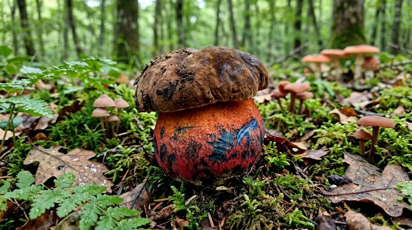
Neoboletus erythropus (Dotted Stem Bolete)
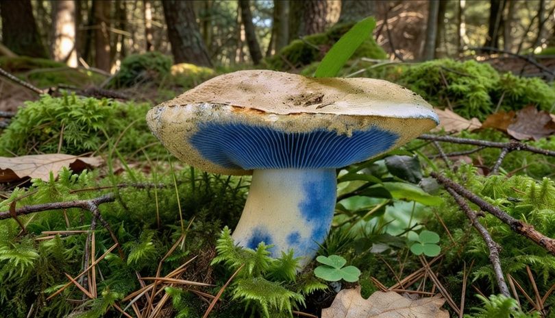
Gyroporus cyanescens (Cyanescent Bolete)
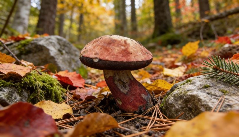
Suillellus luridus (Dusky Bolete)
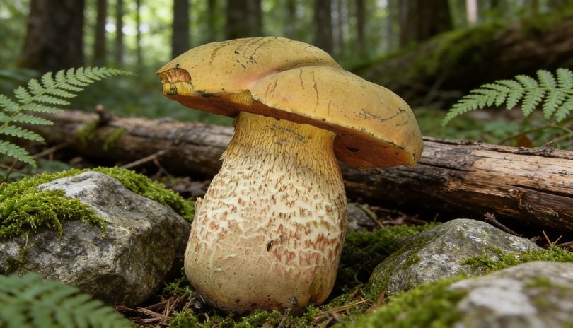
Boletus calopus (Bitter Bolete)
Boletus erythropus var. discolor and related species
for prized species like shiitake, pleurotus, and ganoderma. Guaranteed quality, fast shipping.
Blue staining in psilocybin species
Mushrooms that turn red, orange, and purple
Red staining in boletes and russulas
Yellow staining: the case of Agaricus
How to test the yellow staining of an Agaricus in the field
Purple and lilac staining in Cortinarius
Mushrooms that blacken: the case of Lactarius and Agaricus
The case of Lactarius: milk and oxidation
Species Fresh latex color Color after 5-10 minutes Edibility Lactarius deliciosus Carrot orange Remains orange, then green Edible Lactarius deterrimus Pale orange Turns olive green Edible (inferior) Lactarius salmonicolor Salmon orange Does not change significantly Edible Lactarius indigo Indigo blue Remains blue-green Edible Lactarius controversus White Remains white Toxic Lactarius necator White, then gray Blackens Toxic Lactarius scrobiculatus White, then yellow Turns yellow Toxic Lactarius piperatus White, does not change Remains white Edible (after preparation)
Lactarius deliciosus: the saffron milk cap
Lactarius indigo: the blue milk cap
Blackening of Russula nigricans and relatives
Blackening of Agaricus: button mushrooms and cultivated mushrooms
Chromatic shifting and identification: practical field guide
How to perform the staining test
Diagnostic table: staining as an identifying characteristic
Observation Probable diagnosis Recommended action Rapid and intense blue staining, brown cap, red tubes Neoboletus erythropus or related species Edible cooked, verify absence of red net on stem (which would indicate B. luridus) Slow blue-green staining, orange latex Lactarius deliciosus or deterrimus Edible, L. deterrimus less prized Yellow staining at stem base, phenol odor Agaricus xanthodermus Do not collect, causes gastroenteritis Red → black slow staining (hours), flesh without latex Russula nigricans Not edible or of poor quality, verify other characteristics No staining, white latex does not change color Many possibilities; from other characteristics Continue with other morphological characteristics Electric blue staining on pressure, floury odor Possible Gyroporus cyanescens Edible, rare species Progressive purple-gray staining, fibrous cap Cortinarius sp. (possible) Do not collect, dangerous genus
The limits of staining as a diagnostic characteristic
Chemical reagents used for artificial staining
Why staining is not an indicator of toxicity
Mushrooms that stain and are edible
Mushrooms that don't stain and are dangerous
Critical conclusion: chromatic shifting is an identifying characteristic, not a food safety test. The only safe way to consume wild mushrooms is certain identification of the species by a qualified expert.
Mushrooms change color during cooking: what happens in the kitchen
Thermal reactions in mushrooms
Deactivation of oxidative enzymes
Maillard reaction
Thermal degradation of preformed pigments
Why some mushrooms blacken during cooking
How to preserve mushroom color in the kitchen
Implications for home and professional cultivation
Staining as a quality indicator in mycelium
Staining in cultivated fruiting bodies
Strategies to reduce post-harvest staining in cultivation
Strategy Mechanism Efficacy Applicability Cold chain (2-4°C) Slows enzymatic reactions High Post-harvest and transport Modified atmosphere (MAP) Reduces O₂ available for oxidation Very high Industrial packaging Washing with 0.1% citric acid Lowers pH, inhibits tyrosinase Medium Pre-packaging UV-C treatment Inactivates surface enzymes, reduces microbial load Medium-high Packaging lines Selection of low-tyrosinase strains Fewer oxidative enzymes in genotype High (long term) Breeding and varietal selection Harvest at optimal maturity stage More compact flesh, less mechanical damage Medium Harvest management
Staining as a research tool in breeding programs
Scientific research and pharmacological potential of fungal pigments
Fungal pigments and antioxidant activity
Antibiotic and antifungal potential
Fungal pigments and neuroprotection
Blue staining chemistry as a biotechnological model
Medicinal mushrooms and controlled oxidation: the case of Ganoderma
How to preserve mushrooms by slowing oxidation
Collection: preventing mechanical damage
Short-term storage (refrigerator)
Long-term storage
Drying
Freezing
Preserved in oil and vinegar
Powder and extracts
Tools and resources for the amateur mycologist
Reference books for Italian mycology
Apps and digital resources
Italian mycological associations
FAQ: most frequent questions about mushroom color change
Why do mushrooms change color when they are cut?
Which mushroom turns blue when cut?
Is a mushroom that changes color poisonous?
Why does my cultivated button mushroom turn dark brown if I leave it in the air?
What causes the blackening of mushrooms during cooking?
How can the color change of mushrooms be slowed down?
Do cultivated mushrooms change color like wild ones?
Is the colored latex of lactarius (Lactarius) dangerous?
Is it true that lemon prevents mushrooms from blackening?
Why is fresh porcini different from dried porcini in color?
Data, statistics, and research on fungal staining: an updated overview
Scientific production on fungal staining
Year Number of publications (Scopus, search "mushroom color change") Main discoveries 2000-2005 ~45 Preliminary identification of staining substrates in boletes 2006-2010 ~80 Cloning and characterization of Boletales laccases 2011-2015 ~135 Clarification of the molecular mechanism of blue staining (variegatic acid) 2016-2020 ~200 Biotechnological applications of fungal laccases; pharmacological potential of pigments 2021-2024 ~290 Genomics of staining, pigment biosynthesis, nutraceutical applications
Statistics on mushroom foraging and poisoning in Italy
Parameter Average annual data Italy Notes Habitual mushroom foragers ~3.5 million MIPAAF data 2022 Reported poisoning cases/year 600-900 Source ISS Deaths from mushroom poisoning/year 3-10 Variable by year Species responsible for most deaths Amanita phalloides (~90%) Does not stain when cut Species responsible for most gastroenteritis Agaricus xanthodermus (~30% of cases) Stains yellow Months with highest incidence September-November Coincides with porcini season Mushrooms change color, and we learn to read nature
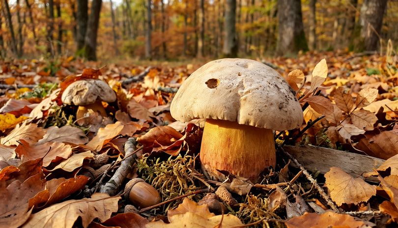
Continue your journey in the world of mushrooms The fungal kingdom is a universe in continuous evolution, with new scientific discoveries emerging every year on their extraordinary benefits for intestinal health and general well-being. From today on, when you see a mushroom, you will no longer think only of its flavor or appearance, but of all the therapeutic potential it contains in its fibers and bioactive compounds. ✉️ Stay connected - Subscribe to our newsletter to receive the latest studies on: Nature offers us extraordinary tools to take care of our health. Mushrooms, with their unique balance between nutrition and medicine, represent a fascinating frontier that we are only beginning to explore. Continue to follow us to discover how these extraordinary organisms can transform your approach to well-being.