


The use of agar agar represents one of the most fundamental and essential techniques in modern mycology, offering mycologists and cultivators a solid and sterile medium for the propagation and study of fungal mycelium. This article delves into the most advanced methodologies for maximizing the potential of this extraordinary polysaccharide, with detailed recipes, sterilization protocols, and inoculation techniques that guarantee success in multiplying the most prized fungal strains. Through this comprehensive guide, we will explore every aspect of preparing and using agar-based culture media, providing scientific data, detailed statistics, and tested procedures to optimize results in mushroom cultivation at both professional and amateur levels.
Agar agar has revolutionized microbiology and mycology since its introduction in the nineteenth century, offering an elegant solution to the problem of cultivating microorganisms on solid substrates. Derived from red algae of the genera Gelidium and Gracilaria, this natural polymer possesses unique properties that make it ideal for creating culture media: it is metabolically inert, resistant to degradation by most microorganisms, and forms a stable gel at temperatures ranging from 35°C to 100°C. The sol-to-gel transition occurs at about 32-40°C, allowing the addition of heat-sensitive nutrients without compromising the structure of the medium. A thorough understanding of these physicochemical properties is essential to fully exploit the potential of agar in mycelial propagation. The history of agar agar in mycology begins at the end of the 19th century, when the German microbiologist Walther Hesse, on the suggestion of his wife Fanny, began using it as an alternative to animal gelatin which liquefied at incubation temperatures. This casual but brilliant discovery allowed Robert Koch to develop the first solid culture media for bacteria, laying the foundation for modern microbiology. In the field of mycology, agar quickly became the standard for the isolation and purification of fungal strains, gradually replacing empirical methods based on natural substrates such as grain, sawdust, or wood. The standardization of agar-based culture media allowed significant advances in fungal taxonomy, the study of fungal physiology, and the development of controlled cultivation techniques. Agar agar is a complex polymer composed mainly of agarose and agaropectin. The gelation mechanism is physical rather than chemical: the polymer chains form a three-dimensional structure that traps water molecules through hydrogen bonds. The typical concentration for mycological applications ranges from 1.5% to 2% by weight, which corresponds to 15-20 grams per liter of medium. At this concentration, the resulting gel has a firm but not excessively rigid consistency, allowing both mycelial growth and the ability to perform transfers without damaging the fungal structure. The gelation temperature of agar (approx. 32-40°C) is significantly lower than its melting temperature (approx. 85°C), creating a wide hysteresis zone that greatly facilitates laboratory procedures. Preparing culture media with agar agar requires precision and attention to detail, as the composition of the medium directly influences the growth rate, mycelial morphology, and expression of the fungus's genetic characteristics. There are numerous recipes, from the simplest to the most complex formulations, each with specific advantages and applications. In this section, we will explore the fundamental recipes and their variants, with precise instructions on concentrations, pH, and preparation protocols. The choice of appropriate medium depends on the fungal species, the purpose of the culture (isolation, preservation, inoculum production), and the available environmental conditions. The Malt Extract Agar (MEA) medium is the most widely used culture medium in mycology for its versatility and excellent support for the growth of a wide variety of fungi. The classic formulation requires 20 grams of barley malt extract, 15-20 grams of agar agar, and 1 liter of distilled water. The malt extract provides a balanced source of simple and complex carbohydrates, amino acids, vitamins, and essential minerals for mycelial development. The pH of the finished medium normally settles between 5.0 and 5.5, ideal for most saprophytic and mycorrhizal fungi. The standardization of MEA has allowed significant comparisons between different strains and species, facilitating the work of identification and classification. Preparing MEA requires attention to sterility and precision in measurements. Start by accurately weighing 20g of barley malt extract and 15g of high-purity agar agar, preferably in fine powder for better solubility. Add these components to 1 liter of distilled water in a beaker or Pyrex flask, stirring with a magnetic stirrer while heating over medium heat. Bring to a gentle boil, continuing to stir until the components are completely dissolved (about 5-10 minutes). It is essential to avoid vigorous boiling to prevent sugar caramelization and thermal degradation of nutrients. After complete dissolution, the medium is sterilized in an autoclave at 121°C for 15-20 minutes, then cooled to 45-50°C before pouring into sterile Petri dishes. The Potato Dextrose Agar (PDA) medium is particularly appreciated for cultivating fungi that require complex carbon sources and a more articulated nutritional profile than simple MEA. The traditional preparation involves infusing 200g of fresh, peeled, and cubed potatoes in 1 liter of distilled water for 30-60 minutes at 60-70°C, followed by filtration and the addition of 20g of dextrose and 15g of agar agar. The potatoes provide starch, minerals, and growth factors that stimulate the development of particularly vigorous and compact mycelium. PDA is particularly indicated for basidiomycetes such as Pleurotus ostreatus, Lentinula edodes, and Agaricus bisporus, which show faster and more structured growth on this substrate. The basic PDA can be significantly improved by adding specific supplements that enhance its performance for particular applications. For lignicolous fungi and particularly demanding species, the addition of 2g/L of yeast extract is recommended, providing B vitamins and essential amino acids. To promote sporulation of recalcitrant strains, supplementation with 0.5g/L of thiamine hydrochloride (vitamin B1) can be decisive. In cases of recurring bacterial contamination, the addition of 50mg/L of streptomycin sulfate or 100mg/L of ampicillin creates a selective medium that inhibits bacterial growth without negatively affecting the fungal mycelium. For fungi requiring particularly acidic pH, such as some Aspergillus and Penicillium, it is possible to add tartaric acid to bring the pH to 3.5-4.0. While standard media like MEA and PDA are suitable for a wide range of fungi, particular species or specific applications require specialized formulations that meet the unique metabolic needs of each genus. In this section, we will explore media optimized for specific genera, with compositions that maximize growth, promote in vitro fruiting, or support particular enzymatic activities. Customizing the culture medium represents a powerful tool for optimizing yield, controlling mycelial morphology, and inducing specific metabolic pathways of interest. Cultivating pure mycorrhizal fungi on agar represents a particular challenge, as these organisms have evolved a metabolic dependence on the host plant. Media for mycorrhizal fungi must contain complex carbon sources like starch or cellulose rather than simple sugars, which can inhibit growth or induce abnormal mycelial forms. An effective formulation includes 10g of potato starch, 2g of monobasic potassium phosphate, 1g of magnesium sulfate, 0.5g of calcium chloride, 15g of agar agar, and 1L of distilled water. The pH should be adjusted to 5.8-6.2 to simulate soil conditions. The addition of small amounts of humic compounds (0.1-0.5g/L) can further mimic the natural root environment. To stimulate the growth of mycorrhizal fungi in the absence of the host plant, the medium can be integrated with extracts from the roots of specific host plants. The procedure involves collecting young roots of the host plant, their surface sterilization with 70% ethanol and 0.5% sodium hypochlorite, followed by homogenization in sterile distilled water and filtration. The extract is added to the culture medium at varying concentrations from 1% to 5% before sterilization. Alternatively, the addition of phytohormones such as auxins (indole-3-acetic acid 0.1-1mg/L) or cytokinins (kinetin 0.01-0.1mg/L) can partially mimic the chemical signals of the host plant. These approaches have been shown to significantly increase the growth of Tuber melanosporum, Cantharellus cibarius, and other economically important mycorrhizal fungi. Sterility is the critical factor determining success or failure in agar cultivation. Even the perfectly formulated culture medium will be useless if contaminated by competing microorganisms. In this section, we will analyze the most effective sterilization protocols, from standard to advanced techniques, with particular attention to contamination prevention and emergency management. Understanding the microbiological principles behind sterilization allows for adapting protocols to specific needs and solving problems that could otherwise compromise entire culture batches. Autoclaving represents the most reliable sterilization method for agar-based culture media. The physical principle is based on the use of superheated steam at high pressure, which allows reaching temperatures above 121°C necessary to destroy all vegetative forms and the most resistant spores. The standard cycle involves exposure to 121°C for at least 15-20 minutes at a pressure of 1 atm above atmospheric pressure. It is essential to consider that the effective sterilization time begins only when all material in the autoclave has reached the target temperature, which may require several additional minutes depending on the container volume. For media particularly rich in organic material or with high spore content, it is advisable to extend the sterilization time to 25-30 minutes. The effectiveness of autoclaving is strongly influenced by the volume and geometry of the containers used. For standard Petri dishes (90-100mm diameter), it is recommended not to exceed 25-30mL of medium per plate, while for laboratory flasks the optimal volume is 200-250mL for a 500mL flask or 400-450mL for a 1000mL flask. These volume/surface ratios ensure efficient heat transfer during sterilization and adequate oxygenation during mycelial growth. Containers with flat bottoms and straight walls allow more homogeneous heat distribution compared to those with curved bottoms or irregular shapes. It is essential to leave caps slightly loose during autoclaving to allow gas expansion and prevent container implosion during cooling. Inoculation of culture media represents the most delicate phase of the entire process, where the mycelium is transferred under conditions of absolute sterility. Inoculation techniques influence not only the colonization speed but also the strain viability and genetic purity of the culture. In this section, we will explore standard and advanced inoculation methods, with particular attention to minimizing contamination risk and optimizing mycelial development. Mastery of these techniques is essential for maintaining pure strains, preventing culture degeneration, and ensuring reproducible results over time. Transferring from a mother culture to new agar plates represents the most common operation in mycelial propagation. The correct aseptic technique involves using an inoculation loop previously sterilized in a flame which, after cooling, is used to take a fragment of mycelium from the mother colony. The sample should be taken from the active growth zone, typically 1-2 cm from the edge of the colony, where the mycelium is young, vital, and genetically stable. The fragment, ranging in size from 2x2 mm to 5x5 mm, is then deposited in the center of the new plate and lightly pressed to ensure good contact with the agar surface. For particularly slow-growing species, multiple points on the same plate can be inoculated to reduce colonization times. The choice of tissue to sample for transfer significantly influences the characteristics of the new culture. The mycelium located immediately behind the growth front presents maximum metabolic activity and genetic stability, while the older central zones may show signs of senescence or mutations. For species that form rhizomorphs, the sample should include at least one complete rhizomorph segment, as these specialized structures contain genetically complementary nuclei necessary for vigorous growth. In fungi that produce clamp connections, it is useful to verify the presence of these structures under the microscope before transfer, as an indicator of a healthy and genetically stable dikaryotic mycelium. Regular microscopic verification of mother cultures is crucial for early identification of signs of degeneration or contamination. Mycelial growth on agar depends not only on the composition of the culture medium but also on a series of environmental parameters that must be carefully controlled to maximize results. Temperature, humidity, lighting, and ventilation interact in complex ways influencing growth speed, mycelial morphology, and the expression of specific characteristics. In this section, we will analyze in detail the effect of each parameter and provide guidelines for optimizing culture conditions based on fungal species and specific objectives. Temperature is one of the most critical factors for mycelial growth on agar. Each fungal species has an optimal temperature range that can vary considerably, from 4-10°C for psychrophilic fungi like Hericium erinaceus to 35-40°C for thermophiles like Aspergillus fumigatus. Most cultivated fungi grow optimally between 20 and 28°C. It is important to note that the optimal temperature for vegetative growth may differ significantly from that required for fruiting. For temperate species, the application of thermoperiods (cyclic temperature variations) can stimulate more vigorous growth and induce the formation of reproductive structures. Precise temperature control allows for synchronizing cultures and optimizing production times. The relative humidity in the incubation environment directly affects the evaporation rate from the agar, which in turn influences nutrient concentration and mycelial morphology. Too low relative humidity (below 60%) causes excessive evaporation, resulting in medium concentration and possible growth inhibition. Conversely, too high humidity (above 90%) promotes condensation on plate walls, increasing contamination risk and creating anaerobic microenvironments. The optimal humidity for most agar cultures is between 75% and 85%. To maintain constant conditions, incubators with humidity control can be used, or simpler systems such as closed containers with saturated saline solutions that stabilize relative humidity at specific values. Despite precautions, agar cultures can encounter various problems that compromise mycelial growth or favor the development of contaminants. The ability to promptly identify these problems and intervene correctly is crucial for maintaining healthy and productive cultures. In this section, we will analyze the most common problems, their causes, and practical solutions, also providing preventive strategies to minimize the risk of failure. A systematic approach to problem-solving allows transforming failures into learning and improvement opportunities. Contaminations represent the most frequent and frustrating problem in agar cultures. Correct identification of the contaminant is the first step to determine its origin and prevent recurrences. Bacterial contaminations typically manifest as slimy, shiny colonies with uniform color, while molds form fluffy or powdery colonies of various colors. Yeasts produce pasty and creamy colonies. Cross-contamination from other fungal cultures can be particularly insidious, as the contaminant might be morphologically similar to the desired mycelium. In case of contamination, it is essential to immediately isolate the affected culture, identify the possible source (air, tools, operator, medium), and review sterilization and aseptic work protocols. Air quality in the work environment is a critical factor in preventing contaminations. Laminar flow cabinets represent the gold standard for aseptic work, providing a constant flow of air filtered through HEPA filters that remove particles and microorganisms. The efficiency of a laminar flow cabinet depends on proper maintenance (regular filter replacement, integrity testing) and appropriate use (disinfected work surfaces, limited movements during operations). In the absence of a laminar flow cabinet, still air boxes can be used which, although less efficient, significantly reduce contamination risk by creating a semi-protected environment. Regular disinfection of the work environment with sodium hypochlorite, ethanol, or hydrogen peroxide is essential to maintain acceptable sterility standards. Long-term preservation of agar cultures is essential to maintain the viability, genetic stability, and specific characteristics of fungal strains. Different methods offer different trade-offs between duration, complexity, and reliability. In this section, we will explore the most effective techniques for culture preservation, from simple methods for hobbyists to cryogenic protocols for reference collections. The choice of appropriate method depends on available resources, frequency of culture use, and the importance of the preserved genetic material. Refrigeration preservation represents the simplest method for medium-term storage of agar cultures. Fresh cultures grown on Petri dishes can be stored at 4°C for 3-6 months, depending on the species and medium composition. To further extend the duration, impoverished media (with lower nutrient concentration) can be used, slowing down the mycelium's metabolism. Cultures stored in refrigeration require periodic transfers to fresh medium to maintain viability. This method presents the risk of mutation accumulation and genetic drift due to repeated subculturing, but remains widely used for its simplicity and accessibility. The cryopreservation in liquid nitrogen (-196°C) represents the most effective method for long-term preservation of fungal cultures. At these temperatures, all metabolic and chemical processes are virtually suspended, allowing for practically unlimited preservation without genetic alterations. The standard protocol involves suspending fragments of mycelium or spores in a cryoprotective solution (generally 10% glycerol or 5% dimethyl sulfoxide), followed by controlled freezing (1°C/minute) to -80°C and subsequent transfer to liquid nitrogen. Cryoprotectants prevent the formation of intracellular ice crystals that would damage cellular structures. This method is particularly indicated for reference collections, mutant strains, and precious genetic material, guaranteeing stability for decades. Precise quantification of mycelial growth on agar is essential for evaluating the effectiveness of different culture media, environmental conditions, or experimental treatments. Various parameters can be measured, each providing specific information about the fungus's growth characteristics. In this section, we will explore the most common methodologies for growth measurement, appropriate statistical analysis techniques, and result interpretation. The quantitative approach allows objective comparisons between strains, systematic optimization of protocols, and a deeper understanding of fungal physiology. Measuring colony diameter represents the simplest and most widespread method for quantifying mycelial growth on agar. Using a caliper or image analysis system, it is possible to track the increase in colony diameter over time, generating growth curves that can be analyzed to determine kinetic parameters such as lag time, exponential growth rate, and stationary phase. For non-perfectly circular colonies, it is common practice to measure two perpendicular diameters and calculate the average. This method is particularly useful for rapid screenings and comparisons between different strains or conditions, although it does not provide information on mycelial density or biomass. Computerized image analysis techniques offer a more sophisticated approach for characterizing mycelial growth. Using specialized software like ImageJ, it is possible to quantify not only the diameter but also the colony area, mycelial density, and texture parameters. The analysis of images acquired at regular intervals allows building high temporal resolution growth curves and identifying complex growth patterns. More advanced techniques like Fourier analysis can characterize the structural complexity of the mycelium, while color analysis can reveal information about produced metabolites. These approaches, although more complex, provide a complete characterization of colony morphology and development. Agar cultivation techniques are not limited to simple fungus propagation but find advanced applications in fields such as genetic engineering, selective breeding, and mutant screening. In this section, we will explore how specialized culture media and agar selection techniques can be used to develop strains with enhanced characteristics, to study fungal genetics, and for biotechnological applications. These applications represent the frontier of applied mycology, with implications for agriculture, medicine, and industry. Agar culture media can be designed to select specific mutants or strains with desired characteristics. The addition of antibiotics, antifungal agents, or metabolic inhibitors creates selective conditions that allow only resistant strains to grow. Similarly, the omission of specific nutrients (minimal media) can select strains capable of synthesizing those compounds. For screening strains with improved enzymatic activity, chromogenic substrates that produce visible signals when hydrolyzed can be incorporated into the medium. These techniques are widely used in breeding programs to develop strains with higher yield, pathogen resistance, or ability to degrade complex substrates. The production and regeneration of protoplasts on agar represents a fundamental technique for fungal genetic engineering. Protoplasts are fungal cells deprived of the cell wall through enzymatic treatment, thus becoming permeable to external DNA. After transformation with the desired genetic construct, the protoplasts are regenerated on agar with osmoprotectants (usually 0.6M MgSO4 or sucrose) that prevent osmotic lysis. Regeneration media are typically enriched with nutrients to support cell wall reconstitution and resumption of cell division. Transformants are then selected on media containing antibiotics or other selective markers. This technique has allowed the development of edible fungus strains with improved characteristics, such as greater pathogen resistance or optimized nutritional content. The use of agar agar in mycelium multiplication represents a mature but continuously evolving technology, with new applications and improvements regularly emerging. From basic formulations to highly specialized media, from simple transfer techniques to cryogenic preservation protocols, agar remains the foundation of practical mycology. Future research directions include the development of chemically defined media for precise metabolic studies, the integration of sensors into culture media for real-time monitoring of growth parameters, and the optimization of sustainable formulations that reduce environmental impact. A thorough understanding of the principles and techniques presented in this article provides the basis for exploring these emerging frontiers and contributing to the advancement of mycological science. The future of agar cultivation appears promising, with several lines of research that could revolutionize current practices. The development of alternative hydrogels with improved properties (greater transparency, thermal stability, adjustable mechanical properties) could overcome some limitations of traditional agar. Microfluidics applied to fungal cultures allows high-throughput screening of thousands of conditions simultaneously, significantly accelerating research. The integration of omics techniques (genomics, transcriptomics, proteomics, metabolomics) with agar growth studies promises to unveil the complex metabolic networks regulating fungal development. These advancements, combined with the traditional techniques described here, open new possibilities for understanding and exploiting the potential of fungi in agriculture, medicine, and biotechnology.Agar Agar in mycology: history and fundamental principles
The historical origins of Agar use in mycology
The gelation mechanism: a detailed scientific explanation
Concentration (%) Gel consistency Recommended applications Melting temperature (°C) Gelation temperature (°C) 1.0-1.2 Soft Cultures of watery fungi, mobility studies 82-85 32-35 1.5-1.8 Medium (Standard) General culture, isolation, preservation 85-88 34-38 2.0-2.5 Firm Long-term cultures, strains producing lytic enzymes 88-92 36-40 3.0+ Very Firm Special applications, resistance to desiccation 92-95 38-42 Basic Agar Agar culture media recipes
Malt extract Agar (MEA): the standard medium par excellence
Detailed MEA preparation: step-by-step procedure
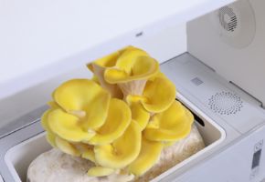
Malt concentration (g/L) Growth speed Mycelial density Rhizomorph formation Recommended applications 10 Slow Low Rare Strain preservation, sporulation induction 20 Medium Medium Moderate General use, propagation 30 Fast High Frequent Inoculum production, slow strains 40+ Very Fast Very High Abundant Research, biomass production PDA (Potato Dextrose Agar): the nutritive medium for demanding fungi
Advanced PDA Variants: Enrichments and Modifications
Specialized culture media for specific fungal genera
Media for Mycorrhizal Fungi: simulating root symbiosis
Integration with root extracts and plant hormones
Fungal genus Optimal medium composition Ideal pH Growth temperature (°C) Colonization time (days) Agaricus MEA + 2g/L yeast extract + 1g/L hydrolyzed casein 7.0-7.5 24-26 10-14 Pleurotus PDA + 5g/L rice bran + 0.5g/L manganese sulfate 6.0-6.5 25-28 7-10 Lentinula MEA + 3g/L wood extract + 1g/L glycine 5.5-6.0 22-25 12-16 Tuber Starch 10g/L + KH2PO4 1g/L + oak root extracts 3% 6.2-6.8 18-20 20-30 Advanced sterilization and preparation protocols
Autoclaving: physical principles and critical parameters
Volume management and container geometries
Volume per container (mL) Heating time (min) Sterilization time at 121°C (min) Cooling time (min) Contamination risk (%) 10-20 5-8 15 10-15 2-5 21-50 8-12 20 15-25 5-8 51-100 12-18 25 25-40 8-12 101-200 18-25 30 40-60 12-18
Advanced inoculation techniques and mycelium transfer
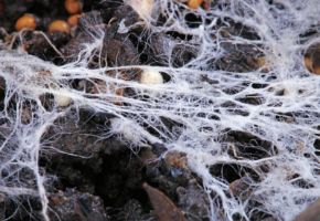
Transfer from mother culture: fundamental aseptic techniques
Tissue selection and genetic stability
Optimization of environmental parameters for growth on Agar
Temperature control: thermal gradients and thermoperiods
Relative humidity and evaporation control
Fungal species Optimal temperature (°C) Relative humidity (%) Photoperiod (light/dark hours) Optimal CO2 (ppm) Agaricus bisporus 24-26 80-85 0/24 2000-5000 Pleurotus ostreatus 25-28 75-80 12/12 1000-2000 Lentinula edodes 22-25 80-85 16/8 1000-1500 Ganoderma lucidum 26-30 85-90 12/12 1500-3000
Troubleshooting common problems in Agar cultures
Identification and management of contaminations
Air quality control and laminar flow cabinet
Long-term preservation techniques for Agar cultures
Refrigeration preservation: techniques and limitations
Cryopreservation in liquid nitrogen: the gold standard
Preservation method Estimated duration Post-preservation viability (%) Equipment costs Technical complexity Refrigeration (4°C) 3-6 months 80-95 Low Low Mineral Oil 1-2 years 70-90 Low Medium Lyophilization 5-10 years 40-70 Medium High Cryopreservation (-196°C) 10+ years 80-95 High High
Statistical analysis and measurement of mycelial growth
Colony diameter measurement and kinetic analysis
Image analysis and computerized techniques
Advanced applications: genetic engineering and breeding on Agar
Mutant selection and screening on selective media
Protoplasts and genetic transformation on Agar
Agar Agar: indispensable for professional cultivators
Research perspectives and future developments