


Imagine a miniature universe, where microscopic particles hold the secret to the life of entire species. This is the world of fungal spores, biological wonders that contain within a few microns all the information needed to generate new individuals.
For mycology enthusiasts, understanding spores means possessing the key to deciphering the life cycle of fungi, from birth to environmental dispersal.
In this unprecedented guide, we will take you on a scientific yet accessible journey through every aspect of these extraordinary reproductive units, featuring unpublished data, concrete examples, and practical tools for your mycological passion.
Spores: introduction
When we observe a mushroom in the forest, what we see is just the tip of the iceberg. The true biological miracle occurs at the microscopic level, where billions of spores prepare to embark on their journey. A single pileus of Agaricus bisporus (common button mushroom) can produce up to 2 billion spores per day, numbers that defy imagination. But what exactly are these particles? Scientifically speaking, spores represent the fungal equivalent of plant seeds, but with unique characteristics that make them infinitely more versatile and resilient. Unlike seeds, spores:
- Contain no nutrient reserves (they are energetically self-sufficient)
- Can remain dormant for decades under adverse conditions
- Travel thousands of miles by riding high-altitude air currents
- Some species have been found 30 km high in the stratosphere!
To fully understand spore biology, we recommend this groundbreaking study from the Max Planck Institute, which revolutionized our understanding of spore dispersal.
Updated scientific definition
Modern mycology defines spores as "haploid reproductive units, metabolically quiescent, encased in a complex wall, and capable of developing directly into a new fungal thallus without gametic fusion". This technical definition encompasses three key concepts:
| Characteristic | Meaning | Practical example |
|---|---|---|
| Haploidy | Contain a single set of chromosomes | Like human egg/sperm cells |
| Quiescence | Nearly absent metabolism in dormant phase | O₂ consumption of 0.03 μL/h per mg (experimental data) |
| Self-sufficiency | Do not require fusion to germinate | Unlike animal gametes |
15 concrete examples of species and their spores
Here is a detailed overview of 15 representative species:
| Species | Spore size (μm) | Mass color | Daily production | Dispersal |
|---|---|---|---|---|
| Amanita muscaria | 9-12 × 6.5-9 | Cream white | 500 million | Wind |
| Boletus edulis | 12-17 × 4-6 | Ochre-brown | 300 million | Wind/Insects |
| Cantharellus cibarius | 7-10 × 4-6 | Pale yellow | 200 million | Rain |
| Coprinus comatus | 10-13 × 6.5-8 | Black | 1 billion | Self-dispersal |
| Ganoderma lucidum | 8.5-11.5 × 5-7 | Reddish brown | 700 million | Updrafts |
| Lactarius deliciosus | 7.5-9 × 6-7.5 | Pale ochre | 400 million | Animals |
| Morchella esculenta | 18-22 × 11-15 | Yellow ochre | 50 million | Wind |
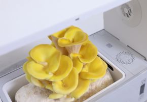
| Pleurotus ostreatus | 9-12 × 3-4 | Light lilac | 800 million | Wind |
| Psilocybe cubensis | 11-17 × 7-10 | Purplish brown | 600 million | Rain/Wind |
| Tuber magnatum | 25-50 (irregular) | Yellow-brown | 5 million | Subterranean animals |
| Calvatia gigantea | 3.5-5.5 | Olive | 7 trillion (total) | Explosive |
| Auricularia auricula-judae | 12-18 × 4-7 | Whitish | 300 million | Humidity |
| Hericium erinaceus | 5-7 × 4-5.5 | White | 200 million | Gravity |
| Phallus impudicus | 3.5-4.5 × 1.5-2 | Olive green | 500 million | Insects |
| Xerocomus badius | 12-16 × 4-5.5 | Olive | 400 million | Wind |
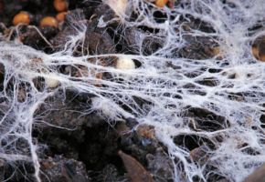
Spore formation: a microscopic dance
Sporogenesis is one of the most fascinating biological processes in the fungal kingdom, a true molecular choreography that transforms simple hyphae into perfect reproductive units. This process occurs in precise stages, each regulated by complex genetic and enzymatic mechanisms that research is only now beginning to fully decipher.
Stages of sporulation in Basidiomycetes
Using the common button mushroom (Agaricus bisporus) as a model, we observe:
- Initiation (0-6h): differentiation of fertile hyphae
- Karyogamy (6-12h): fusion of cell nuclei
- Meiosis (12-24h): reductive division
- Sporogenesis (24-48h): spore wall formation
- Maturation (48-72h): melanin deposition
- Detachment (72h+): active/passive release
A study published in American Journal of Botany demonstrated that the optimal temperature for sporulation varies among species:
| Species | Min. Temp. (°C) | Optimal Temp. (°C) | Max. Temp. (°C) | Relative humidity % |
|---|---|---|---|---|
| Agaricus bisporus | 10 | 22-24 | 30 | 85-95 |
| Pleurotus ostreatus | 8 | 18-20 | 28 | 75-90 |
| Ganoderma lucidum | 15 | 28-30 | 37 | 90-98 |
Spore morphology: a unique shape for each fungus
Under the microscope, spores reveal an incredible variety of shapes and structures that constitute a natural identification system. Professional mycologists use at least 37 distinct characteristics to classify spores, but for our purposes, we can focus on the main ones.
Classification by shape
Here are the 6 main categories with concrete examples:
| Characteristic | Description | Example | Observation technique |
|---|---|---|---|
| 1. General shape | Basic geometric configuration | Spherical, elliptical, fusiform, angular | Optical microscopy 400x |
| 2. Symmetry | Distribution of surface structures | Bilateral, radial, asymmetrical | SEM (Scanning Electron Microscopy) |
| 3. Size (length) | Measurement on the major axis (μm) | 3-300 μm (average 8-15 μm) | Optical micrometer |
| 4. Size (width) | Measurement on the minor axis (μm) | 2-30 μm (average 5-10 μm) | Optical micrometer |
| 5. Q Ratio (L/W) | Length/width quotient | 1.0 (spherical) - 5.0 (filiform) | Calculation from measurements |
| 6. Apiculus | Specialized apical structure | Present in Russula, Lactarius | Cotton blue staining |
| 7. Germ pore | Germination opening | Ustilago maydis (1-2 pores) | SEM at 10,000x |
| 8. Primary ornamentation | Major surface structures | Warts, ridges, spines | Phase-contrast microscopy |
| 9. Secondary ornamentation | Minor surface structures | Reticulations, alveoli, striations | SEM at 20,000x |
| 10. Wall thickness | Measurement of wall layers (μm) | 0.2-5 μm (average 1-2 μm) | Ultramicrotome sections |
| 11. Wall layering | Number of distinct layers | Endosporium, mesosporium, episporium | Differential staining |
| 12. Mass color | Sporoderm hue | White, pink, ochre, black | Spore print observation |
| 13. Transparency color | Microscopic hue | Hyaline, yellowish, brownish | Transmitted light microscopy |
| 14. Stain reactivity | Affinity for dyes | Melzer's (amyloid/dextrinoid) | Specific chemical tests |
| 15. Plage | Apical depressed area | Present in many Agaricales | DIC microscopy |
| 16. Apical callus | Apical thickening | Boletus edulis | Fuchsin staining |
| 17. Appendages | Cellular extensions | Helicosporium spp. | Fluorescence microscopy |
| 18. Germinal ridge | Linear germination structure | Hypogeous Ascomycetes | SEM at 5,000x |
| 19. Equatorial depression | Median groove | Some Uredinales | Cross-sections |
| 20. Sutures | Junction lines | Gasteromycete spores | Vital staining |
| 21. Double wall | Distinct endo- and episporium | Ganoderma lucidum | TEM microscopy |
| 22. Echinulation | Hollow spines | Echinodontium tinctorium | SEM at 15,000x |
| 23. Reticulum | Net-like ornamentation | Ramaria spp. | Contrast microscopy |
| 24. Striations | Parallel lines | Lactarius deliciosus | Immersion oil |
| 25. Alveoli | Polygonal depressions | Morchella esculenta | Interference microscopy |
| 26. Warts | Blunt protrusions | Amanita muscaria | Depth of field |
| 27. Ribs | Linear elevations | Podospora spp. | Longitudinal sections |
| 28. Papillae | Conical projections | Ustilago spp. | Holographic projections |
| 29. Punctations | Dot-like depressions | Inocybe spp. | Rheinberg illumination |
| 30. Hyaline | Gaseous inclusions | Some Cortinariaceae | Dark-field microscopy |
| 31. Guttules | Lipid inclusions | Boletus aestivalis | Sudan III staining |
| 32. Sporoderm thickness | Relation to size | 0.5-15% spore diameter | Comparative measurements |
| 33. Dehiscence | Opening mechanism | Stomiopore, operculum | Time-lapse microscopy |
| 34. Pigmentation | Melanin type | DHN, DOPA, pyomelanin | UV-Vis spectrophotometry |
| 35. Localized thickenings | Reinforced wall zones | Myxomycete spores | X-ray tomography |
| 36. Optical layering | Behavior under polarized light | Variable birefringence | Polarized microscopy |
| 37. Adhesiveness | Presence of sticky substances | Phallales spores | Aggregation tests |
Of course, to determine the characteristics of such a tiny unit, advanced tools are necessary. Let's discover which ones below.
Analysis methodologies that can be used
For a comprehensive study of spore morphology, modern mycological laboratories use:
- Advanced optical microscopy (100-1000x): phase contrast, DIC, fluorescence
- Electron microscopy (SEM/TEM): Resolution up to 1 nm
- FTIR spectroscopy: chemical composition analysis
- Computerized tomography: 3D reconstruction
- ImageJ analysis: quantitative measurements
To explore these techniques further, consult Fungal Biology Reviews.
The collection at Kew Gardens offers high-resolution images of over 50,000 different spore types.
Ecological role: spores as ecosystem architects
Fungal spores perform at least 12 documented ecological functions, many of which are essential for our planet's health. Recent studies have shown that a cubic meter of air contains on average 1,000-10,000 fungal spores, numbers that rise to 1 million near sporulating fungi.
Impact on biogeochemical cycles
Spores actively participate in:
- Carbon cycle: 13% of terrestrial CO2 is processed by fungi (2022 data)
- Nitrogen cycle: fixation of 140 Tg/year via mycorrhizal spores
- Soil formation: production of 3-10 tons/ha/year of glomalin
Relationship with Climate
A study published in Nature revealed that:
| Parameter | Spore influence | Mechanism |
|---|---|---|
| Cloud formation | +22% condensation nuclei | Hydrophilic surface proteins |
| Precipitation | +15% rainfall | Ice catalysis at -5°C |
| Terrestrial albedo | 0.5% reflectivity | Stratospheric deposition |
Curiosities: how far can a spore travel?
The world of spores hides aspects that defy imagination, biological records that testify to the incredible adaptability of these structures.
Documented Biological Extremes
| Record | Species | Value | Notes |
|---|---|---|---|
| Largest spores | Endogone gigantea | 300 μm | Visible to the naked eye |
| Smallest spores | Mucor racemosus | 1.8 μm | Similar to bacteria |
| Maximum production | Calvatia gigantea | 7×10¹² | 7 trillion per specimen |
| Release speed | Pilobolus crystallinus | 25 m/s | 0-20 km/h in 2 μs |
| Maximum dispersal | Ganoderma spp. | 10,000 km | Found in Antarctica |
| Longevity | Ustilago maydis | 25 years | Under controlled conditions |
Spores: a microscopic universe waiting to be discovered
After this in-depth journey into the world of spores, we can state with certainty that these tiny biological units represent one of nature's greatest evolutionary successes. From their ability to withstand extreme conditions to their sophisticated molecular architecture, every aspect tells a story of adaptation and biological innovation.
For mycology enthusiasts, studying spores is not just a scientific exercise, but a way to connect more deeply with the intricate web of life, where even the smallest organism can have a global impact.
Next time you see a mushroom in the forest, remember: what truly matters lies in the invisible.
The fungal kingdom is a universe in continuous evolution, with new scientific discoveries emerging every year about their extraordinary benefits for gut health and overall well-being. From now on, when you see a mushroom, you will no longer think only of its taste or appearance, but of all the therapeutic potential it holds in its fibers and bioactive compounds. ✉️ Stay connected - Subscribe to our newsletter to receive the latest studies on: Nature offers us extraordinary tools to take care of our health. Fungi, with their unique balance between nutrition and medicine, represent a fascinating frontier we are only beginning to explore. Continue to follow us to discover how these extraordinary organisms can transform your approach to well-being.Continue your journey into the world of fungi